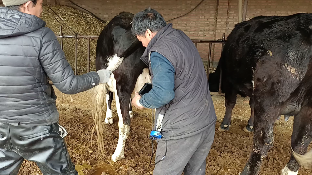
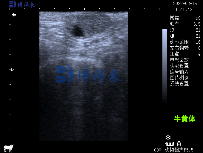
The ultrasound characteristics of the corpus luteum in cows are of great clinical significance. Using a veterinary ultrasound machine, the boundaries of the corpus luteum can be clearly observed. Its echo is messy and weaker than the ovarian stroma. The corpus luteum is usually gray, oval, and clearly separated from other structures on the ovary. In particular, the sunken corpus luteum, the so-called cystic corpus luteum, can also be accurately analyzed by ultrasound.

The formation of the corpus luteum originates from the hypertrophy and luteinization of the granulosa cells and theca cells in the inner wall of the follicle. Its development speed is extremely fast, and its diameter can usually reach 10 mm within 2 days. On the 7th to 8th day of the menstrual cycle, the corpus luteum reaches its maximum size, and its diameter can fluctuate between 20 and 25 mm. However, shortly after ovulation, the newly developed corpus luteum cannot be detected. It can only be detected by a Veterinary ultrasound machine within 2 to 4 days after ovulation.
At a frequency of 5 MHz, the corpus luteum is always visible from the beginning of corpus luteum development to the end of the estrus period. Although the size of the corpus luteum of different ages is small and therefore cannot be used for specific disease diagnosis, ultrasound examination is still one of the main means for veterinary ultrasound machines to detect ovarian corpus luteum. It should be noted that the echo characteristics cannot accurately distinguish between the corpus luteum of pregnancy and the normal corpus luteum.

Ultrasound manifestations of cystic corpus luteum
The internal depression of the cystic corpus luteum is usually oval or round and is almost always located in the inner half of the organ. The maximum size of its cavity is generally between a few millimeters and 1.5 centimeters. The echo is similar to that of the follicle, and sometimes slight reflections in the intracavitary fluid can be seen. With the help of veterinary ultrasound machines, cystic corpora luteum can be accurately distinguished from ovarian follicles.
Growth cycle of corpus luteum and ultrasound detection
Veterinary ultrasound machines play an indispensable role in animal reproduction management. Through ultrasound examination, veterinarians can accurately determine the number of corpora luteum on the ovary and their development. The pits in the cystic corpus luteum usually reach their maximum width between the 8th and 10th days of the menstrual cycle, and then their frequency and size will gradually decrease.
Differences in Corpus Luteum among Different Animals
The size of the corpus luteum is closely related to the progesterone concentration, and the echogenicity of the corpus luteum of buffalo is similar to that of cows, but smaller in size (10 to 14 mm) and with a lower incidence of local depressions. Although veterinary ultrasound is more accurate than rectal palpation in determining follicular development, it is still difficult to distinguish between developing and degenerating corpora lutea.
With the help of veterinary ultrasound, veterinarians can efficiently monitor the development and changes of the corpus luteum. Ultrasound examination provides a powerful tool for animal husbandry, both in reproductive management and disease diagnosis.
tags: Veterinary B-ultrasound machineVeterinary B-ultrasound


