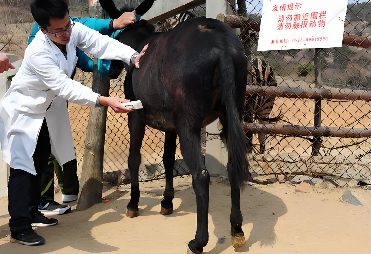
Due to the much smaller size of mini donkeys compared to regular donkeys, it is not possible to fully introduce hands and arms into the rectum for ultrasound testing like detecting regular donkeys. Therefore, specialized ultrasound probes are required for rectal testing of mini donkeys. Firstly, use gloves and lubricated hands to empty the feces from the anus with three fingers. Then, apply the coupling agent onto the probe, which helps to safely introduce the probe into the appropriate examination position and provide good contact between the probe and the rectal wall to improve imaging of the adjacent uterus and ovaries.

Mini Donkey Rectal Examination
If there is resistance or fecal interference with the transmission and reception of ultrasound when slowly pushing the donkey with the B-ultrasound probe, it should be removed and 300 milliliters of warm soapy water can be administered for enema. Send the animals to the recycling area and restart the inspection after the animals have excreted their feces. In practice, less than 10% of rectal examinations require this procedure. Before introducing the B-ultrasound probe into the rectum, the donkey is further lubricated by applying lubricant externally.
It initially advanced 45 degrees upwards through the anal sphincter to allow the donkey pelvis to tilt. About 4 to 6 inches inside the rectum, the bladder will be visible at the 6 o'clock position (bottom). This is a convenient landmark for visualizing the uterus, which is located outside the bladder. As the probe advances, the main body of the Y-shaped uterus is considered a linear structure. When the probe is advanced and rotated to the 12 o'clock and 14 o'clock positions approximately 5 to 7 inches after insertion, a cross-sectional view of the uterine angle can be obtained. The probe may need to be moved in and out or slightly rotated to locate the left and right corners for examination (see the following image: longitudinal transrectal ultrasound image of the miniature donkey uterus during estrus).
Longitudinal transrectal ultrasound images of miniature donkey uterus during estrus period
Once the minimum diameter of the corresponding angle is found, the probe will rotate to the 3 o'clock or 9 o'clock position of the insertion depth to locate the ovary. Again, a slight rotation, insertion, or removal of the probe may be necessary to view the entire ovary (see image below: ultrasound views of the two ovaries of a miniature donkey during estrus).
Donkey pregnancy detection using B-ultrasound machine
Sometimes, due to the presence of feces between the probe and the rectal wall, it may not be possible to examine one side or the other. Sometimes a probe can be used to gently rotate feces for a clearer view. On certain days, due to interference in the movement of the intestine between the rectum and reproductive organs, it is impossible to locate the ovaries or fully examine the uterus. It is best to stop the detection and try again. If the animal strains at any time during the inspection process, it should be terminated immediately. The risk of rectal injury is very low, and careful and slow examination eliminates this possibility.
High definition donkey B-ultrasound pregnancy tester training
tags: ultrasound probesB-ultrasound machineultrasound machine


