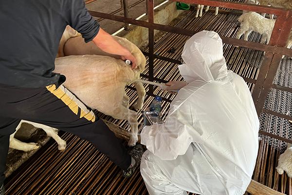
Animal B-ultrasound can detect the subcutaneous fat and/or maximum depth of the longest muscle and/or the surface area of the longest muscle between the 12th and 13th ribs in sheep, which are commonly used indicators for predicting sheep carcass. Although ultrasound measurements of the longest muscle width and area have significant predictive factors (P<0.15) in a few cases, they generally do not contribute significantly to the prediction accuracy of GR, lean meat production, and saleable meat production. There is a high correlation between the longest muscle area determined by the digital board and the longest muscle area determined by image analysis. The longest muscle area determined based on carcass measurements is an important predictor of beef meat production. If there is a method to accurately measure the longest muscle area using ultrasound, sheep may also be the same. One possible reason for the decrease in measurement accuracy using animal ultrasound is the presence of several small muscles, such as the multifidus dorsi and costal longus, located near the longest muscle. In lambs lacking a layer of intermuscular fat, these smaller muscles may appear as part of the longest muscle in ultrasound images.

Sheep's backfat eye muscle area
The animal B ultrasound machine was used to carry out ultrasound measurement on four parts of the left side of the sheep: the total tissue depth between the 11th and 12th ribs, 11cm outside the spine and parallel to it (longitudinal measurement, flat gel pad; fat depth (FD12us) and LM depth (LD12us) between the 12th and 13th ribs perpendicular to the midline of the body (transverse measurement, curved gel pad); Fat depth (FD3Tus) and LM depth (LD3Tus) between the 3rd and 4th lumbar vertebrae, perpendicular to the spine (transverse measurement, curved gel pad); Fat depth (FD3Lus) and LM depth (LD3Lus) between the 3rd and 4th lumbar vertebrae, parallel to the midline of the body (longitudinal measurement, flat gel pad). The same operator performed ultrasound measurements throughout the entire experimental process. Capture images at each site and measure immediately using the device's cursor. For lateral measurement, the probe is placed perpendicular to the spine to capture the entire lamb chops, the maximum height of the LM, perpendicular to the surface, and the depth of fat beyond this height are allocated to muscle and fat depth, respectively. The longitudinal measurement parallel to the backbone captured the image of LM along its length. In this case, the muscle depth corresponds to the maximum height between the transverse processes, and the fat depth is the fat coverage at that muscle depth. Skin depth is included in all ultrasound fat measurements because the tissue is not easily distinguishable from the fat layer. The skin layer is very thin (2.5 to 3.0 millimeters), and the differences between animals are minimal. Our measurements and analysis of skin thickness around 110 days in this experiment confirmed these observations (3.5 ± 0.4 millimeters; data not shown)
tags: animal B-ultrasound


