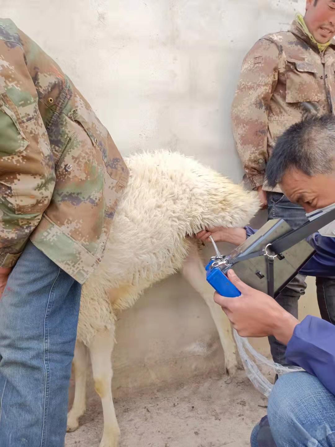
The benefits of diagnosing goat pregnancy have been widely accepted, and the use of a sheep ultrasound scanner equipped with a 5 MHz linear array or sector transducer is suitable for goat uterine ultrasound examination. The sector system requires less skin surface contact, which can reduce the scanning time required for each animal. When pregnancy exceeds 100 days and the uterus reaches a more anterior abdominal position, a 3.5 MHz transducer may be required to penetrate to the desired depth.
High definition sheep ultrasound machine

The transabdominal ultrasound scan of goat uterus is preferably performed when the animal is in a standing position. It is best to conduct the inspection when the goat is standing on the milking stable or milking stand, as the investigator can easily apply the transducer in a standing position without bending over. In addition, several goats can be tied together and given some food to make them stand more quietly. Depending on the breed and coat, it may be necessary to scrape off a portion of the right abdominal wall in front of the breast, approximately 6 x 12 centimeters. This improves the contact between the transducer and the skin, thereby enhancing the image quality. Ultrasound examination is best performed from the right side, as filling the rumen from the left side can hinder correct observation of the uterus. However, if there are doubts about the quality or interpretation of the image from the right side, scanning from the left side can sometimes provide better results. After applying the coupling gel, place the transducer on the abdominal wall. If from the 50th to the 100th day of pregnancy, the uterus will immediately appear on the screen. If this is not the case, the ultrasound beam must be directed towards the pelvic entrance. Subsequently, the tail of the abdomen was explored by rotating the linear array transducer along its longitudinal axis.
Ultrasound examination of the uterus can also be performed through the transrectal approach, where the transducer is inserted into the rectum through an extension rod. In this way, sometimes the ovaries can be seen. This technology has been used to evaluate the response of goats to superovulation in embryo transfer procedures.
When it is necessary to scrape the abdominal wall, the transrectal route can be more time-saving than the transabdominal route for goat uterine ultrasound examination. By preventing the transducer from coming into close contact with the rectal wall, fecal consistency may pose some difficulties during transrectal scanning. When inserting the extension rod into the rectum, ewes usually have less cooperation compared to ewes, which may lead to rectal injury or even perforation, similar to perforation caused by other pregnancy diagnostic methods such as rectal abdominal palpation. According to reports, palpation of the rectum and abdomen in goats can sometimes lead to miscarriage.
tags: B-ultrasound machinesheep ultrasound machineultrasound machine


