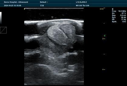
In the world of veterinary medicine, early diagnosis and accurate detection of health issues in animals are key to ensuring their well-being and improving their quality of life. Veterinary ultrasonic detection is a non-invasive, highly effective diagnostic tool that has revolutionized how veterinarians assess and treat a wide variety of animal health conditions. From detecting internal injuries to monitoring pregnancies, ultrasonography has become an indispensable part of modern veterinary care. This article explores the importance of Veterinary ultrasound, its applications, benefits, and why it has become a go-to diagnostic tool in veterinary practices worldwide.

What is Veterinary Ultrasound Detection?
Veterinary ultrasound detection refers to the use of ultrasound technology to create images of an animal’s internal organs and tissues. This technology uses high-frequency sound waves that are emitted from an ultrasound probe, which then bounce off internal structures and are returned to the probe. These reflected sound waves are processed into an image that can be viewed in real-time on an ultrasound screen.
Unlike traditional X-rays, ultrasound does not use radiation, making it a safer, non-invasive option for diagnosing health conditions in animals. The real-time imaging provided by veterinary ultrasound allows veterinarians to get an accurate view of soft tissues, such as muscles, tendons, and organs, making it invaluable for diagnosing a range of conditions.
Applications of Veterinary Ultrasound
1. Pregnancy Detection and Reproductive Health
One of the most common uses of veterinary ultrasound is for pregnancy detection in animals, particularly in cattle, horses, dogs, and cats. Early pregnancy detection helps veterinarians monitor the health of the fetus and provide early interventions if needed. With ultrasonic imaging, veterinarians can observe the developing fetus, assess the health of the uterus and ovaries, and check for complications such as multiple pregnancies, fetal death, or ectopic pregnancies.
In addition to pregnancy checks, veterinary ultrasound plays a crucial role in evaluating reproductive health. For example, it is used to detect ovarian cysts, uterine infections, and other reproductive conditions in both female and male animals.
2. Internal Organ Health
Veterinary ultrasound allows veterinarians to examine internal organs, such as the liver, kidneys, spleen, and heart, to detect abnormalities that might indicate underlying health issues. Abdominal ultrasounds are used to assess the size, shape, and structure of these organs, identifying potential issues like tumors, cysts, infections, or congenital defects.
For example, if a dog shows signs of gastrointestinal distress, an abdominal ultrasound can help diagnose conditions such as gastrointestinal blockages, inflammatory bowel disease (IBD), or liver disease.
3. Musculoskeletal System Evaluation
Ultrasound is also invaluable for examining the musculoskeletal system of animals, especially in large animals like horses and cattle, where joint, bone, and soft tissue injuries are common. Ultrasound imaging allows veterinarians to visualize tendons, ligaments, and muscles to assess for tears, strains, or damage.
This is particularly useful for athletic animals (such as racehorses or working dogs) to detect soft tissue injuries, such as tendonitis, ligament damage, and muscle tears that could affect their performance and mobility.
4. Cardiac Ultrasound (Echocardiography)
Echocardiography, or cardiac ultrasound, is a specialized form of ultrasound that examines the heart and blood vessels. It is used to assess heart function, check for heart murmurs, valvular disease, arrhythmias, and other cardiac abnormalities. This is particularly important in aging pets or animals with suspected heart conditions, as it allows veterinarians to measure heart size, blood flow, and overall heart health in real-time.
5. Cancer Detection
Veterinary ultrasound is frequently used to detect signs of cancer or tumors in animals. It is an effective tool for identifying soft tissue tumors that may not be visible through traditional imaging methods like X-rays. Ultrasound allows veterinarians to visualize the size and location of tumors, and to determine whether they have spread to surrounding tissues or organs. It is often used in conjunction with biopsy or aspiration to help confirm a diagnosis of cancer.
Benefits of Veterinary Ultrasonic Detection
1. Non-Invasive and Safe
Unlike some diagnostic procedures, such as X-rays or CT scans, ultrasound does not involve radiation, making it a safe and non-invasive procedure for animals of all ages. This is particularly important for pregnant animals or young animals, where radiation exposure can be harmful to developing embryos or growing animals.
2. Real-Time Imaging for Immediate Diagnosis
The ability to view real-time imaging means that veterinarians can make quick, informed decisions about a pet’s or livestock’s health. This is especially valuable when diagnosing time-sensitive conditions, such as intestinal blockages, fetal distress, or acute injuries. The ability to visualize movement and structure in real-time helps veterinarians guide their next steps in diagnosis and treatment.
3. Cost-Effective and Time-Saving
Ultrasound is often more cost-effective and less time-consuming compared to other imaging technologies like CT scans or MRIs. In many cases, ultrasound can provide the necessary diagnostic information without the need for expensive, time-intensive procedures. It is also easier to conduct on-site or in the field, especially for farm animals or large animals like horses and cattle.
4. Enhanced Diagnostic Accuracy
Ultrasound offers high levels of accuracy, allowing veterinarians to identify a wide range of conditions that might be missed by physical examination or other diagnostic methods. Its ability to visualize soft tissues provides more detailed and comprehensive diagnostic data, allowing for early intervention and preventative care.
5. Minimal Discomfort for the Animal
Since ultrasound is non-invasive, animals experience minimal discomfort during the procedure. The process typically requires only the application of a special gel to the skin, which is much less stressful compared to more invasive procedures like surgery or biopsy. As a result, animals can often remain calm and comfortable during the scan, making it an excellent choice for routine check-ups and health monitoring.
How Veterinary Ultrasound Works
The ultrasound machine works by emitting high-frequency sound waves through a transducer (probe), which is placed on the animal’s body. These sound waves travel through the body and are reflected by tissues and organs, creating an image. The image is then displayed on a monitor for the veterinarian to analyze. Different tissues reflect sound waves differently, so the machine uses these echoes to build detailed images of the internal structures.
Common Types of Veterinary Ultrasound:
- Abdominal Ultrasound: Used for imaging the abdominal organs like the liver, kidneys, and intestines.
- Cardiac Ultrasound: Focuses on the heart and blood vessels.
- Musculoskeletal Ultrasound: Examines muscles, ligaments, tendons, and joints.
- Reproductive Ultrasound: Used for pregnancy detection and reproductive health monitoring.
tags:


