When your furry friend isn’t feeling well, veterinarians often rely on advanced tools to peek inside their bodies without surgery. One such tool is Veterinary ultrasound—a safe, non-invasive imaging technology that uses sound waves to create real-time pictures of organs, tissues, and blood flow. Whether it’s checking a cat’s heart, monitoring a dog’s pregnancy, or diagnosing liver issues in horses, ultrasound has become a cornerstone of modern animal care. Let’s break down how this technology works, step by step.

The Basics of Sound Wave Imaging
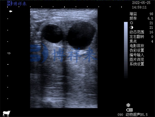
Veterinary ultrasound machines send high-frequency sound waves (inaudible to humans or animals) into the body using a handheld probe. These waves bounce back when they hit tissues, fluids, or organs, much like how a bat navigates using echolocation. The returning echoes are captured by the probe and translated into visual images by specialized software. For example, fluid-filled areas like the bladder appear dark, while denser tissues like bones show up brighter. This process happens in real time, allowing vets to observe moving structures like a beating heart or intestinal activity .
Key Components of an Ultrasound Machine
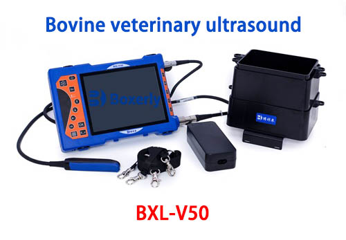
A typical ultrasound system includes three main parts: the transducer (probe), processing unit, and display screen. The transducer is the most interactive component—vets move it over the animal’s shaved and gel-coated skin to capture images. Modern probes are designed for different purposes. For instance, a convex probe might scan a dog’s abdomen, while a microconvex probe could examine a rabbit’s tiny organs. The processing unit uses algorithms to sharpen images and reduce noise. Take BXL’s systems: their Fusion THI 2.0 technology enhances resolution, making it easier to spot subtle abnormalities in tissues .

From Waves to Diagnostic Images
Once echoes are detected, the machine’s software maps their intensity and location to build a 2D or 3D image. Advanced features like Doppler mode add color to visualize blood flow direction and speed—helpful for detecting heart defects or blocked vessels. Some systems, like those with Xbeam technology, improve spatial resolution to reduce shadows, ensuring clearer views of overlapping structures like intestinal loops. Real-time adjustments matter too. If a horse’s thick muscle attenuates the signal, the machine can compensate by boosting weak echoes or suppressing overly bright ones .
Everyday Uses in Animal Health
Ultrasound isn’t just for emergencies. Vets use it routinely for pregnancy checks, guiding needle biopsies, or assessing organ health. For example, a cat with unexplained weight loss might get an abdominal scan to check for kidney disease. In livestock, ultrasound helps monitor fetal development in cows or detect muscle injuries in racehorses. It’s also handy for guiding minimally invasive procedures, like draining fluid from a dog’s cyst. Clinics often pair ultrasound with lab tests (bloodwork, urinalysis) to get a full picture of an animal’s health .

Why Modern Systems Are Game-Changers
Today’s ultrasound devices prioritize ease of use and precision. Features like automated measurements save time—a vet can quickly calculate a heart’s ejection fraction to assess cardiac function. Elasticity imaging, another innovation, maps tissue stiffness to identify tumors. Portable models let vets perform scans in the field, whether at a dairy farm or a pet owner’s home. Systems with nSlice technology even capture split-second movements, like a rabbit’s rapid breathing, without motion blur. These advancements make diagnoses faster and more accurate, improving outcomes for animals worldwide .
tags:


