Ultrasound imaging has revolutionized the way veterinarians around the world diagnose, treat, and monitor tendon injuries in horses. Tendon damage, once considered a career-ending setback for many performance and sport horses, is now routinely assessed and managed with far greater precision. Yet the fundamental question remains: can a horse truly recover from a tendon injury? Drawing on international perspectives—from data-driven Western protocols to integrative approaches in Asia—this article explores tendon anatomy, injury mechanisms, healing biology, rehabilitation strategies, and the critical role of ongoing ultrasound monitoring in returning horses to soundness.
Understanding Equine Tendon Anatomy and Function
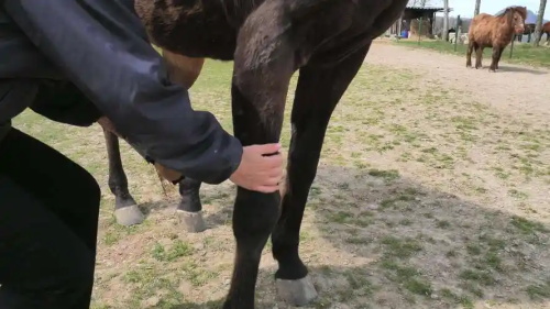
Equine tendons in the distal limb—including the superficial digital flexor tendon (SDFT) and the deep digital flexor tendon (DDFT)—are specialized for energy storage and release. Their unique composition of parallel collagen fibers, proteoglycans, and water gives them both tensile strength and elasticity during high-speed gaits or jumping. However, this same specialization makes them vulnerable to strain injuries:
SDFT (Superficial Digital Flexor Tendon)
Runs from the back of the knee (or hock) down to the pastern, acting as a major shock absorber.DDFT (Deep Digital Flexor Tendon)
Lies deeper in the limb, attaching at the coffin bone and responsible for flexing the toe.
Compared to muscle, tendons have a poor blood supply—up to 50% less vascularity—which hampers their intrinsic healing capacity. In high-performance horses, repetitive loading can produce micro-damage that accumulates, eventually manifesting as a clinically significant lesion.
Types and Grading of Tendon Injuries
Veterinary practitioners classify tendon injuries according to severity and location:
Grade I (Mild Strain)
Minimal fiber disruption, slight swelling, and mild pain on palpation. Often resolves with conservative rest.Grade II (Moderate Strain)
Partial tear of collagen fibers; marked swelling, heat, and palpable defect. Requires extended rehabilitation.Grade III (Severe Rupture)
Complete fiber disruption; severe lameness, hematoma formation, and often a visible “bow” in the tendon. Historically considered career-ending without advanced therapy.
Advanced imaging modalities, especially high-resolution ultrasound, allow precise grading by visualizing fiber alignment, cross-sectional area (CSA), and echogenicity changes.
The Biology of Tendon Healing
Equine tendon healing occurs in three overlapping phases:
Inflammatory Phase (0–7 days)
Platelet-derived growth factors (PDGF), transforming growth factor-β (TGF-β), and macrophages clear debris and recruit fibroblasts.Proliferative Phase (7–21 days)
Fibroblasts synthesize type III collagen in a disorganized matrix; neovascularization occurs.Remodeling Phase (3 weeks–12 months)
Gradual replacement of type III with stronger type I collagen; fibers realign under mechanical load.
Despite this remarkable biology, healed tendon tissue rarely achieves the same biomechanical properties as uninjured tendon—type I collagen fibers may be smaller in diameter and less well organized, predisposing to reinjury.
Clinical and Rehabilitation Strategies
Western Evidence-Based Protocols
In North America and Europe, large-scale studies of retired racehorses and sport horses have informed multimodal rehabilitation:
Strict Stall Rest (first 2–4 weeks)
Minimizes mechanical disruption during the inflammatory phase.Controlled Hand-Walking (weeks 4–8)
Low-impact loading stimulates collagen alignment without overstraining the repair site.Incremental Treadmill or Track Work (weeks 8–16)
Progressive increase in speed and distance to enhance tensile strength.Strengthening Exercises (months 4–12)
Hill work, cavaletti poles, and varied terrain to encourage functional fiber remodeling.
Adjunct therapies such as shockwave therapy, intralesional stem-cell or platelet-rich plasma (PRP) injections, and low-level laser have shown variable success in enhancing healing quality and reducing reinjury rates.
Integrative and Eastern Approaches
In countries like Japan, China, and Korea, traditional medicine often complements Western rehabilitation:
Acupuncture and Electro-acupuncture to modulate local blood flow and reduce inflammation.
Herbal Poultices containing anti-inflammatory botanicals such as rhubarb, turmeric, or cangzhu.
Tui Na Massage and gentle mobilization to maintain joint range of motion without overstressing healing tissue.
While high-quality randomized trials in veterinary integrative therapies remain rare, many practitioners report positive anecdotal outcomes when these are combined judiciously with evidence-based rest and exercise protocols.
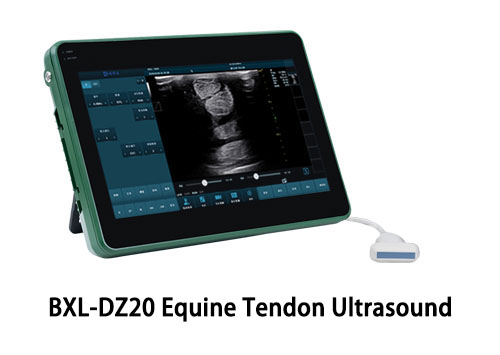
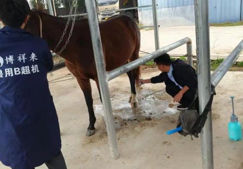
BXL-DZ20 Equine Tendon Rehabilitation Ultrasound
Role of Ultrasound in Monitoring Recovery
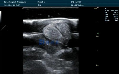
Ultrasonography has become the gold standard for both initial injury assessment and serial monitoring of tendon healing. Key metrics include:
Cross-Sectional Area (CSA):
An acute tendon injury often shows a 30–50% increase in CSA due to fluid and hemorrhage. As healing progresses, CSA should gradually decrease toward baseline.Echogenicity:
Hypoechoic (dark) regions indicate fiber disruption or fluid; progressive return to a more mixed echogenic pattern suggests maturing collagen.Fiber Alignment:
The “feathered” appearance of disorganized fibers should resolve into linear, parallel echogenic bands over months.
International consensus guidelines recommend ultrasound evaluations at:
Injury diagnosis (day 0)
Early proliferative phase (2–4 weeks)
Start of controlled exercise (8–12 weeks)
Mid-rehabilitation (4–6 months)
Return-to-work clearance (9–12 months)
Can a Horse Truly Recover?
Success Rates and Return to Performance
Large retrospective studies on Thoroughbred racehorses indicate:
Grade I–II injuries: ~80% return to racing within 6–9 months, with a reinjury rate of 25–35%.
Grade III injuries: ~50% return to any level of work; fewer (<30%) return to previous peak performance.
In sport horse populations (show jumpers, dressage, eventers), careful rehabilitation under an integrated veterinary-physiotherapy program can push return-to-soundness rates above 70% even for moderate lesions.
Risk Factors for Reinury
Common predictors of reinjury include:
Early return to high-speed work (<7 months post-injury)
Insufficient fiber remodeling on ultrasound (persistent hypoechoic areas)
Underlying conformational stresses (long toe/low heel angles, tendon asymmetry)
Incomplete pain management during rehabilitation
Comprehensive management addressing hoof balance, shoeing modifications, and tailored exercise is thus critical for durable recovery.
Global Perspectives: Learning Across Borders
North America & Europe:
Focus on standardized protocols, measurable ultrasound endpoints, and advanced biologic therapies. Clinical research is ongoing into novel scaffolds and growth-factor delivery systems.Asia & Australasia:
Embrace integrative modalities alongside ultrasound-guided interventions. Tendency to incorporate holistic musculoskeletal assessment, including acupuncture meridians and traditional herbal formulations.Emerging Regions (Latin America, Middle East):
Growing access to portable ultrasound systems enables field-based diagnostics and rehabilitation oversight even in resource-limited settings.
Regardless of region, all experienced practitioners agree: tendon injuries demand patience, precise monitoring, and gradual progression. No “quick fix” exists; success hinges on marrying biological healing timelines with mechanical conditioning.
Practical Tips for Optimizing Healing
Optimize Nutrition:
Adequate protein, vitamin C, and copper support collagen synthesis. Omega-3 fatty acids may modulate inflammation.Manage Pain and Inflammation:
NSAIDs judiciously in the acute phase; consider adjuncts like pentosan polysulfate for joint-related inflammation.Maintain General Conditioning:
While the injured limb rests, water treadmill or hand-walk contralateral limbs to maintain cardiovascular fitness.Ensure Farriery Balance:
Heel wedging or therapeutic shoes can offload stress on healing tendons.Involve a Multidisciplinary Team:
Veterinarian, physiotherapist, farrier, and trainer must collaborate on a progressive plan.
Conclusion
Can a horse recover from a tendon injury? The answer today is a qualified “yes.” With a structured rehabilitation timeline, evidence-based therapies, and meticulous monitoring—particularly via ultrasound—many horses can return to their previous athletic level. Yet each case is unique, heavily influenced by lesion grade, individual healing potential, and the rigor of the rehabilitation program.
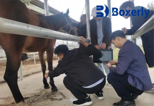
At BXL, we specialize in veterinary ultrasonography solutions that empower equine practitioners to monitor tendon healing in real time. Our BXL-DZ20 portable ultrasound system delivers high-resolution imaging, intuitive software for measuring cross-sectional area and fiber alignment, and wireless data management—so you can track your patient’s progress from the barn aisle to advanced training facilities. Trust BXL to keep every tendon on the path to optimal recovery.
tags:


