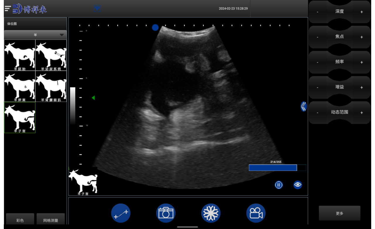
Veterinary ultrasound scans are invaluable diagnostic tools that provide real-time images of a pet’s internal organs and structures. These images can help veterinarians diagnose a variety of health conditions and tailor treatment plans for your furry friend. In this article, we will explore what a veterinary ultrasound scan image is, how to interpret it, and its significance in veterinary medicine.

What is a Veterinary Ultrasound Scan?
A veterinary ultrasound scan utilizes high-frequency sound waves to create detailed images of the inside of an animal’s body. Unlike X-rays, which provide static images, ultrasound allows veterinarians to visualize organs in motion, offering a dynamic view of your pet’s health. The procedure is non-invasive and does not involve radiation, making it a safe option for pets.
How Does an Ultrasound Scan Work?
Preparation: Before the scan, your veterinarian may recommend fasting your pet for several hours to ensure clearer images. This is especially important for abdominal scans.
The Procedure: During the ultrasound, a special gel is applied to your pet’s skin to facilitate sound wave transmission. The veterinarian uses a handheld device called a transducer to send and receive sound waves, capturing the echoes to create images displayed on a monitor.
Image Generation: The ultrasound machine converts the echoes into real-time images, allowing the veterinarian to assess various internal structures, such as the heart, liver, kidneys, and reproductive organs.
Understanding Veterinary Ultrasound Scan Images
Types of Images Produced
Veterinary ultrasound scan images can depict a wide range of structures, including:
Abdominal Organs: Images of the liver, spleen, kidneys, and intestines help diagnose issues such as tumors, cysts, or organ enlargement.
Cardiac Images: Echocardiograms assess heart size, shape, and function, helping to identify conditions like heart disease or congenital defects.
Reproductive Imaging: Ultrasound can confirm pregnancies, assess fetal health, and evaluate reproductive issues in both male and female pets.
Interpreting the Images
While the images generated by an ultrasound scan can be complex, veterinarians are trained to interpret them accurately. Here are some key features they look for:
Size and Shape: Abnormalities in organ size or shape can indicate underlying health issues.
Texture and Consistency: Changes in the texture of organs may suggest inflammation, infection, or tumors.
Fluid Accumulation: The presence of excess fluid in the abdominal cavity or around organs can be a sign of various conditions, including injury or disease.
Importance of Veterinary Ultrasound Scan Images
Early Detection of Health Issues: Ultrasound scans can identify problems before they become serious, allowing for earlier intervention and better outcomes for pets.
Non-Invasive Diagnostic Tool: As a non-invasive procedure, ultrasound minimizes stress and discomfort for pets compared to other diagnostic methods.
Guiding Treatment Plans: The information gathered from ultrasound scan images helps veterinarians tailor treatment plans, ensuring pets receive appropriate care based on accurate diagnoses.
Monitoring Progress: Ultrasound can be used to monitor ongoing health issues, allowing veterinarians to track changes and adjust treatment as necessary.
tags: Veterinary UltrasoundVeterinary Ultrasound ScanVeterinary Ultrasound Scan Image


