


Products Description
")
")
")
")
")
")
")
Tags:Back Fat eyes muscle measuring instrument
Text link:https://bxlultrasound.com/pdt/750.html
Technical parameter

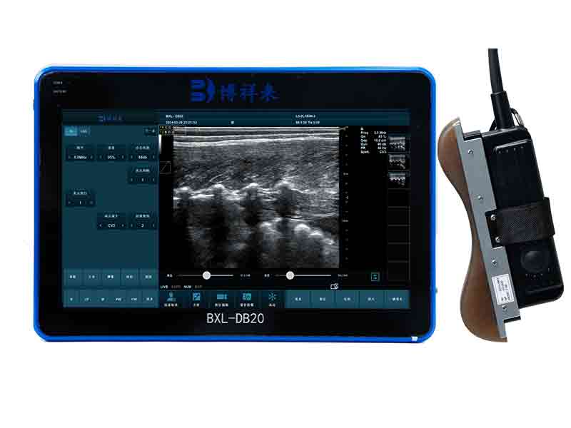
BXL-V60 Eyepiece Vet Ultrasound | Portable Multi-Function Device for Large Farms

BXL-V50 Portable Vet Ultrasound | 8" HD Screen | Flagship Handheld Scanner

BXL-V10Ⅲ Basic USG Device for Livestock & General Veterinary Use | High Cost Performance
1.1. Adaptability: waterproof and dustproof, IP54 waterproof and dustproof level, drop-proof
1.2. Storage and transportation environment: -20°C to +60°C Humidity range: 10%-95%
1.3. Power supply: AC220V±10%, 50Hz±1
1.4. Host size: overall size 400*280*58mm
1.5. Host weight: 6kg
1.6. Host screen size: 15.6-inch touch screen Resolution: 1920*1080
1.7. Host carrying method: handle (hangable), mobile bracket (adjustable angle), can be hung on a mobile cart
1.8. Number of host channels: 64 channels
1.9. Imaging mode: color Doppler imaging system, B, B|B, 4B, B|M (M line position adjustable), M mode, tissue harmonic imaging (THI), pulse inversion harmonic imaging (iTHI), tissue specific imaging (TSI) , convex array extended imaging, linear array deflection/trapezoidal imaging, puncture enhancement, color blood flow (CF), power Doppler (PDI), directional power Doppler (DPDI), pulsed Doppler (PW), continuous Doppler high pulse repetition frequency (CW, HPRF), B+CF color double-width real-time imaging, color M-type, B+PW, B+CF/PDI/DPDI+PW, CW, TDI
1.10, expansion interface: two USB interfaces, 720p HDMI interface, video glasses interface, charging interface
● HDMI, video output; ● USB interface; ● RJ-45/DICOM interface; ● support inkjet, laser, video printer.
1.11, scanning mode: digital beamformer, animal OPU dedicated micro-convex, animal dedicated back fat linear array
1.12. Scanning depth: ≥30cm
1.13. Image processing: multi-beam imaging; high line density scanning (adjustable); 8-segment segment gain TGC control; dynamic range: ≥120 dB; automatic gain control; M scanning speed adjustable; PW scanning speed adjustable; sound power adjustable; controllable frame correlation; Gamma correction; spatial compound imaging adjustable, scanning range adjustable; up/down/left/right/black and white flip; image enhancement, edge enhancement; noise suppression; spot suppression, dynamic focus.
1.14. Image display: 256 grayscale
1.15. Total gain: 0-100 adjustable LGC left and right segment gain 8 segments adjustable TGC upper and lower segment gain 8 segments adjustable.
1.16. Conventional measurement and calculation: distance, depth, area, perimeter, volume, angle, stenosis ratio, ratio, speed, gradient, acceleration, heart rate, etc.
1.17. Animal body mark: ≥24 types Measurement and calculation software package: animal-specific measurement package
1.18. Wireless WiFi connection and Bluetooth connection. Wireless connection to mobile phones and iPads to display images synchronously, with wireless remote control. RFID electronic ear tag scanning
1.19. High-efficiency lithium battery: replaceable Working time: ≥3 hours Battery specification: 10000mAh AC power adapter charging or operation; Output: 14.4V DC 4A
1.20. Image and video storage. Format: AVI, JPG, BMP, PNG, DCM (DICOM), etc. Movie playback function is available. Animal back fat images can be stored in different parts.
1.21. Puncture guide, with puncture line (3 types), scale ruler on the puncture line, center line on the puncture line, and puncture image enhancement processing.
1.22. Built-in professional OPU system program, can set the position and direction of the egg collection needle. There are egg collection needle guide lines (3 types), with scale rulers on the egg collection needle guide lines, center lines on the egg collection needle guide lines, and enhanced image processing when the egg collection needle is punctured.
1.23. Built-in professional back fat eye muscle storage image system, can store back fat eye muscle images according to different parts of different animals for inspection
1.24. Image rotation: 0°, 90°, 180°, 270° rotation; image flip: up and down, left and right
1.25. Scan type: normal, composite expansion, etc.
1.26. Brightness: -50%-50% adjustable Contrast: -50%-50% adjustable
1.27. The host has a stylus; compatible with wireless keyboards and wireless mice
1.28. Optional probe types: animal OPU special micro-convex probe, animal special back fat linear array probe and other probes.
1.29. Image adjustment: Focus adjustment, focus position and number are adjustable. Image frame average adjustment, frame average is adjustable. Spot reduction, with 5 adjustments.
1.30. Image editing: Stored images can be edited. Stored videos can be edited for each frame
1.31. When measuring animal back fat, the back fat measurement and eye muscle depth can be adjusted to be on the same line
1.32. Software version: Animal-specific version
1.33. Patented product: Patent number: ZL 202330405248.6
II. Probe type and parameters:
1. Animal OPU live egg collection probe:
1.1. Frequency 5.0-9.0MHz, non-porous, smooth surface, eagle-beak-shaped integrated micro-convex probe
1.2. Waterproof and dustproof probe
1.3. Number of array elements: 128 array elements
1.4. OPU gun length: 61.5cm (length can be extended according to needs); diameter: ≤2.5cm; weight: ≤700g Cable length: 200cm (standard), customizable length
1.5. Egg collection needle tube: short needle tube, long needle tube, threaded needle tube
1.6. Suitable for short needles, threaded needles, long needles, specific needles, etc.
1.7. Magnetic guide groove (convenient for disinfection and cleaning)
1.8. Egg collection needle tube scale mark, used to control the depth of needle insertion
1.9. There is an egg collection needle guide line when collecting eggs, there is a scale ruler on the egg collection needle guide line, there is a center line on the egg collection needle guide line, and the image is enhanced when the egg collection needle is punctured
1.10. Patented product: Patent number: ZL 202330496819.1
2. Animal back fat eye muscle probe:
2.1. Frequency: 2.5-6MHz 18cm long linear array probe
2.2, Number of array elements: 128 array elements
2.3, Probe line length: 200cm (standard), customizable length
2.4, Probe scanning depth can reach 30cm
2.5, Probe weight: ≤700g
III. Product features of animal OPU live egg collection probe:
1. The eagle-beak-shaped integrated micro-convex live egg collection probe is a product carefully designed according to the clinical practice of the ranch. It has passed the clinical tests of technical experts in the field of animal reproduction for multiple OPU embryo transplantation. It is convenient for clinical operation and has a high egg collection efficiency.
2. The integrated seal of the probe is screw-free, which is convenient for rapid disinfection and cleaning.
3. This probe can be adapted to various types of egg collection needles, which is convenient for different types of customers to adapt to different egg collection needles and save costs.
4. There is an egg collection needle guide line when collecting eggs, a scale ruler on the egg collection needle guide line, a center line on the egg collection needle guide line, and an image enhancement design when the egg collection needle is punctured, which can greatly improve the direction sense and strength control of the needle during egg collection.
5. The diameter of the egg collection gun is very small and can be used for young cows of very young age.
6. High-frequency probe and high-quality Doppler color ultrasound images can observe the situation of follicles below 1mm.
IV. Features of animal backfat eye muscle probe:
1. 18cm long professionally designed backfat eye muscle probe, which can fully test the backfat eye muscles of various animals
2. The probe scanning depth can reach 30cm, which can well present the eye muscles of large animals
3. High-frequency probe and high-quality Doppler color ultrasound images can well identify the measured muscle quality grade
Standard configuration:
Host 1
Backfat eye muscle area probe 1
Silicone pad 1
Lithium-ion battery 1
Adapter 1
Stylus 1
Instruction manual/certificate/warranty card 1 set
Coupon 1 bottle
Trolley case/suitcase: 1
Optional accessories:
Trolley
Wireless keyboard
Wireless mouse
Vacuum pump
Thermostatic bath
