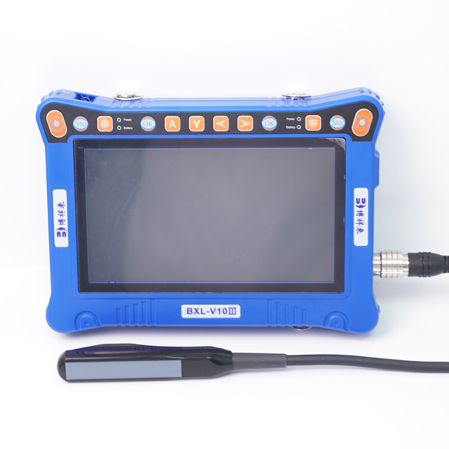
Veterinary ultrasound machines have transformed pet diagnostics, providing veterinarians with the ability to conduct non-invasive examinations and obtain real-time images of a pet's internal structures. To maximize the effectiveness of this advanced technology, it’s essential to follow specific tips and guidelines. This article offers insights into best practices for using veterinary ultrasound machines, ensuring optimal results and improved patient care.

Understanding veterinary ultrasound machines
A veterinary ultrasound machine uses high-frequency sound waves to create detailed images of internal organs and tissues. This non-invasive method is invaluable for diagnosing a wide range of conditions, including tumors, cysts, and cardiac issues. Proper usage of ultrasound machines not only enhances diagnostic accuracy but also improves overall patient outcomes.
Tips for Using Veterinary Ultrasound Machines
Proper Training and Familiarization
- Ensure all staff members operating the ultrasound machine are adequately trained. Familiarity with the device’s features and functions is crucial for obtaining high-quality images and accurate diagnoses.
Choose the Right Transducer
- Different transducers are designed for various applications. select the appropriate transducer based on the examination requirements. For instance, a high-frequency transducer is ideal for superficial structures, while lower frequencies penetrate deeper tissues.
Maintain Equipment Regularly
- Regular maintenance and calibration of the ultrasound machine are essential to ensure optimal performance. Follow the manufacturer’s guidelines for maintenance schedules and address any technical issues promptly.
Use Quality Ultrasound Gel
- Ultrasound gel is crucial for effective imaging. Use high-quality gel to ensure proper sound wave transmission between the transducer and the pet’s skin. Avoid using expired or low-quality gels, as they can affect image quality.
Prepare the Patient Appropriately
- Proper preparation of the pet is essential for obtaining clear images. Ensure the fur is clipped if necessary and apply gel evenly to avoid air pockets. Position the pet comfortably to minimize movement during the examination.
Optimize Imaging Settings
- Adjust the ultrasound machine settings to optimize image quality. This includes selecting the appropriate depth, gain, and focus based on the specific examination area. Familiarize yourself with the machine’s settings to achieve the best results.
Utilize Doppler Imaging When Necessary
- For cardiac assessments or evaluating blood flow, use Doppler imaging capabilities if available. This feature provides valuable information about blood flow dynamics and can help identify potential cardiac issues.
Document Findings Thoroughly
- Keep detailed records of all ultrasound examinations, including images and any notable findings. This documentation is crucial for future reference, follow-up appointments, and communication with pet owners.
Communicate with Pet Owners
- Explain the ultrasound process to pet owners to alleviate any concerns. Discuss findings and potential next steps in a clear and compassionate manner, fostering trust and understanding.
Stay Updated on Advances in Technology
- The field of veterinary ultrasound is continually evolving. Stay informed about new technologies, techniques, and best practices through professional development, webinars, and industry publications.
tags: Veterinary Ultrasound MachineVeterinary Ultrasound


