
Cattle Gestational Sac Ultrasound: How to Identify Early Pregnancy in Cows
Early pregnancy diagnosis is an essential part of modern cattle reproduction management. For veterinarians, cattle breeders, and dairy farm operators, identifying pregnancy at an early stage helps improve breeding efficiency, reduce reproductive delays, and make better herd management decisions.
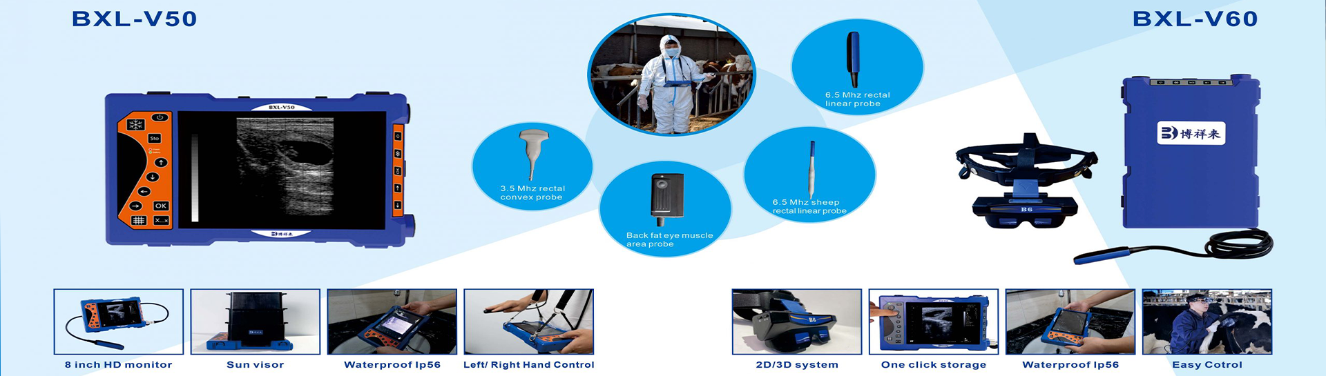
One of the important findings during early bovine pregnancy ultrasound is the gestational sac. By using a professional Veterinary ultrasound scanner, experienced operators can examine the uterus and identify fluid-filled structures associated with early embryonic development.
Understanding how to recognize a cattle gestational sac, when it becomes visible, and which ultrasound equipment is suitable for reproductive examinations can help veterinarians perform more effective pregnancy assessments.
In this article, we explain the role of gestational sac ultrasound in cattle pregnancy diagnosis and introduce professional veterinary ultrasound solutions from BOXERLY.
1. What Is a Gestational Sac in Cattle?
A gestational sac is an early pregnancy-related structure that develops within the uterus after fertilization and implantation.
During early pregnancy, the conceptus develops within the uterine environment. The gestational sac, commonly referred to in practical ultrasound examinations, appears as a fluid-filled structure that can be identified through transrectal ultrasound under suitable examination conditions.
In cattle, ultrasound examination of the gestational sac provides important information about early pregnancy development.
Veterinarians generally evaluate several structures during early pregnancy scanning:
Fluid-filled embryonic vesicle.
Uterine lumen and endometrial structures.
Embryonic development.
Amniotic and associated fetal membranes as pregnancy progresses.
Embryonic heartbeat at an appropriate stage of development.
The appearance and size of these structures change as pregnancy advances.
It is important to distinguish a true gestational sac from other fluid-filled uterine structures, such as uterine fluid associated with reproductive disorders or normal physiological changes.
Therefore, pregnancy diagnosis should be based on a complete ultrasound examination rather than the presence of uterine fluid alone.
2. When Can a Gestational Sac Be Seen in Cattle?
The timing of gestational sac detection depends on several factors, including the cow's reproductive condition, ultrasound equipment, probe frequency, operator experience, and examination technique.
The following timeline provides a general reference for bovine pregnancy ultrasound.
Approximately 18–21 days after insemination
Early pregnancy-related changes may be present, but reliable identification of a gestational sac is challenging. Routine ultrasound confirmation at this stage can be difficult.
Approximately 21–25 days after insemination
In suitable cases, experienced veterinarians may identify an early embryonic vesicle through transrectal ultrasound. However, detection is not guaranteed, and a negative examination should not automatically be interpreted as confirmation that the cow is open.
Approximately 28–30 days after insemination
The gestational sac and embryonic structures are generally easier to identify. This period is more suitable for practical early pregnancy ultrasound examinations.
Approximately 30–35 days after insemination
The embryo becomes more readily visible, and embryonic cardiac activity can often be assessed. This provides additional evidence when confirming early pregnancy.
For better diagnostic reliability, veterinarians should follow appropriate examination protocols and schedule a repeat scan when findings are uncertain.
3. How to Identify a Cattle Gestational Sac Using Ultrasound
Accurate identification of the gestational sac requires appropriate scanning technique and a good understanding of bovine reproductive anatomy.
Transrectal ultrasound is a commonly used method for early pregnancy diagnosis in cattle.
During the examination, the veterinarian introduces a suitable rectal linear probe into the rectum and examines the reproductive tract.
The main examination steps include:
Step 1: Locate the uterus
Identify the uterine body and uterine horns. Examine the reproductive tract systematically to avoid missing early pregnancy-related structures.
Step 2: Examine the uterine lumen
Look for localized fluid-filled structures within the uterine horn. An early embryonic vesicle may appear as a small anechoic or dark fluid-filled area.
Step 3: Evaluate the suspected gestational sac
Assess its location, appearance, and relationship with the surrounding uterine structures.
Step 4: Identify the embryo when visible
As pregnancy progresses, the embryo becomes easier to recognize within the embryonic vesicle.
tags: