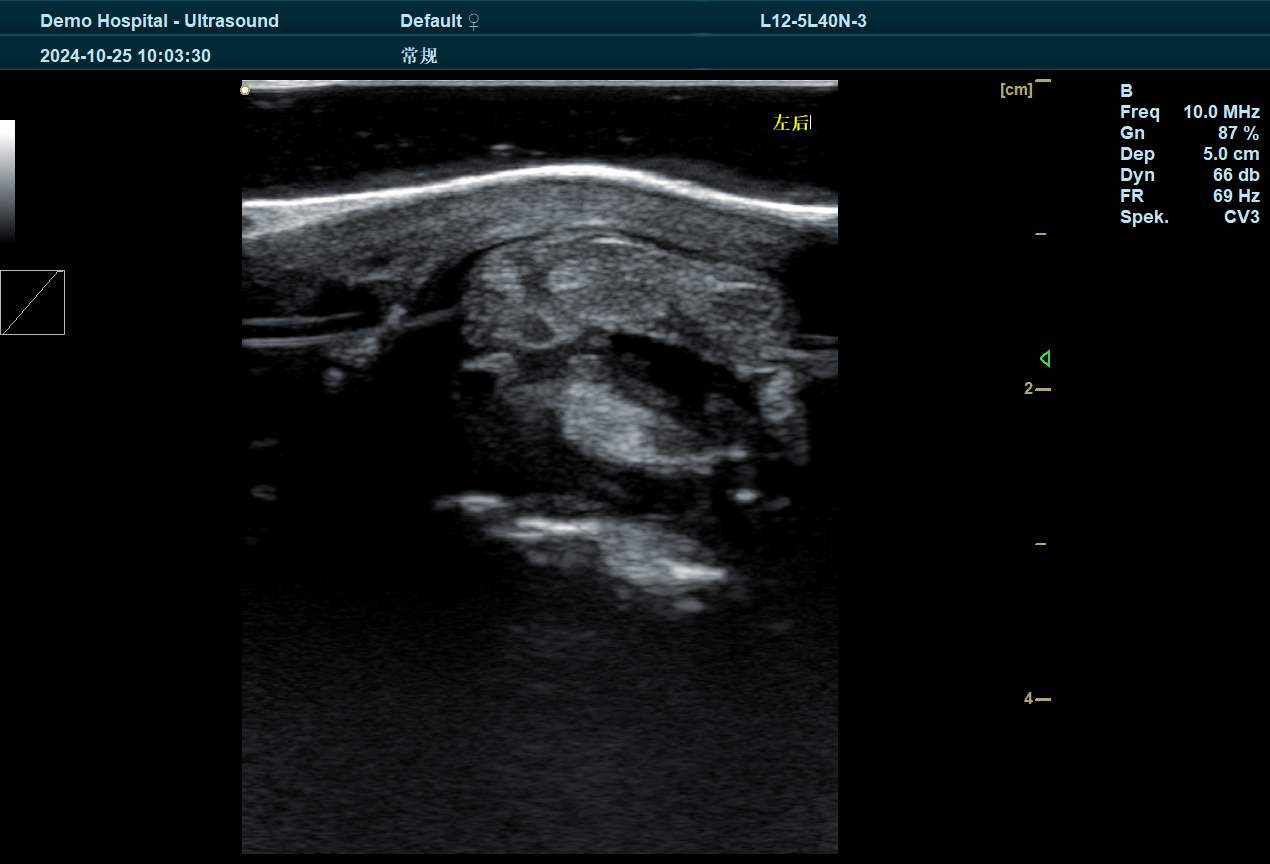
Camel musculoskeletal injuries are common in working camels, particularly those used in agriculture, transportation, and racing. These injuries can range from minor strains to more severe ligament and tendon tears, all of which can affect the camel's ability to perform daily tasks and impact overall health. Early diagnosis and effective treatment are crucial to ensuring quick recovery and preventing long-term complications.

Ultrasound technology, particularly systems like BXL veterinary ultrasound, has become a revolutionary tool in diagnosing and managing camel musculoskeletal injuries. This non-invasive, real-time diagnostic method allows veterinarians to assess the extent of soft tissue injuries, such as muscle strains, tendon ruptures, ligament sprains, and joint problems, with remarkable accuracy.
In this article, we will explore how ultrasound for camel musculoskeletal injuries is enhancing the care of working camels and why it is the preferred diagnostic tool for musculoskeletal problems.
What is Ultrasound for Camel Musculoskeletal Injuries?
Ultrasound for camel musculoskeletal injuries involves using high-frequency sound waves to create detailed images of the internal soft tissues, such as muscles, ligaments, tendons, and joints. The BXL Veterinary ultrasound system is specifically designed to cater to the unique needs of large animals like camels, offering high-resolution imaging that helps veterinarians diagnose injuries early and accurately.
When a camel experiences a musculoskeletal injury, the ultrasound probe is used to send sound waves into the affected area. These sound waves are reflected back to the machine and converted into visual images, allowing the veterinarian to see the extent of the injury, whether it’s a tear, bruise, or inflammation.
Why is Ultrasound Important for Diagnosing Camel Musculoskeletal Injuries?
1. Non-Invasive and Stress-Free
Unlike X-rays or other diagnostic methods, ultrasound is non-invasive and does not require the use of radiation. This makes it an ideal option for diagnosing musculoskeletal injuries in camels, as it is safe for both the animal and the veterinarian. This is especially important for camels that may be sensitive or stressed in clinical settings, as ultrasound allows for a more comfortable and stress-free experience.
2. Real-Time, High-Resolution Imaging
BXL ultrasound provides real-time imaging, which is essential for evaluating musculoskeletal injuries in camels. The ability to see the injury in detail and monitor the healing process over time allows veterinarians to make informed decisions about the treatment plan. Whether it's assessing the severity of tendon damage or detecting swelling, ultrasound offers high-resolution images that help veterinarians spot issues early, leading to better outcomes.
3. Accurate Detection of Soft Tissue Injuries
Soft tissue injuries, such as those affecting muscles, tendons, and ligaments, can be difficult to detect with traditional imaging techniques. Camel tendons and ligaments are especially prone to injuries like sprains, strains, or tears, which may not be visible through an external examination. BXL ultrasound provides clear and precise imaging of these soft tissues, enabling the veterinarian to identify problems such as:
- Tendon tears or ruptures
- Muscle strains and sprains
- Ligament injuries or sprains
- Inflammation or swelling in joints
- Bursitis and fluid buildup
- Bone fractures near tendons or joints
4. Faster Diagnosis and Treatment
Since ultrasound provides immediate results, veterinarians can quickly diagnose the extent of a musculoskeletal injury. Early diagnosis is critical for effective treatment and quick recovery. By visualizing the injury, veterinarians can determine the most appropriate treatment, which could include rest, rehabilitation, or even surgical intervention if necessary.
5. Monitoring Progress and Recovery
Ultrasound is not only useful for initial diagnosis but also for monitoring recovery. Camels that have suffered musculoskeletal injuries often require extended periods of rest and rehabilitation. Regular ultrasound assessments during recovery help veterinarians track the healing process, identify any setbacks, and adjust treatment plans as necessary to ensure the best possible recovery outcomes.
Common Musculoskeletal Injuries in Camels
1. Tendon and Ligament Injuries
Working camels, especially those involved in racing, transport, or carrying heavy loads, are prone to injuries involving their tendons and ligaments. Common tendon injuries include:
- Tendon ruptures: Tears in the tendon fibers, often caused by excessive strain or sudden movements.
- Tendonitis: Inflammation of the tendons, typically due to repetitive strain or overuse.
- Ligament sprains: Damage to the ligament fibers that connect bones to other bones, often caused by twisting or hyperextension.
Ultrasound is highly effective in detecting and assessing the severity of tendon and ligament injuries. The ability to see the exact location and size of tears or inflammation enables veterinarians to make more accurate treatment decisions.
2. Muscle Strains and Sprains
Just like tendons and ligaments, muscles are also susceptible to strain and sprain injuries. These injuries often occur when a camel is overexerted, such as during long-distance travel, heavy lifting, or sudden movements.
Ultrasound can help identify muscle tears, bruising, or swelling that may be causing pain or limiting mobility. Identifying muscle damage early can prevent further injury and facilitate a faster recovery.
3. Joint Injuries
Joint injuries in camels often involve damage to the synovial fluid or cartilage. These types of injuries can lead to joint stiffness, swelling, and lameness. Ultrasound is particularly useful in evaluating joint effusion (fluid accumulation) and detecting soft tissue damage around the joints.
Injuries to the carpal joint, tarsal joint, or fetlock joint are common in camels that are used for heavy labor. Ultrasound can help assess whether joint injuries are superficial or deeper, which is essential for determining the treatment approach.
4. Bursitis
Bursitis occurs when the bursae (fluid-filled sacs that cushion the joints) become inflamed due to overuse or repetitive trauma. Bursitis can cause pain, swelling, and stiffness in the affected joint. Ultrasound can detect bursitis by visualizing the swollen bursa and the surrounding soft tissue, allowing for a more accurate diagnosis and targeted treatment.
5. Bone Fractures
While ultrasound is primarily used for soft tissue evaluation, it can also be useful for detecting fractures or stress fractures in bones that are near tendons and ligaments. Ultrasound can provide clear images of fractures that may not be immediately visible on an X-ray, especially those in the extremities or joints.
How BXL Ultrasound Helps with Camel Musculoskeletal Injuries
1. Portable and Field-Ready
BXL veterinary ultrasound systems are compact, lightweight, and portable, making them perfect for use in the field, especially for working camels. Whether on a farm, at a camel race, or in a remote desert location, veterinarians can perform on-site assessments of musculoskeletal injuries, providing quick diagnoses and immediate treatment plans.
2. Cost-Effective
By using ultrasound as a diagnostic tool, camel owners and veterinarians can avoid the need for costly surgeries or prolonged treatments that may result from undiagnosed or mismanaged injuries. Regular monitoring of injuries using BXL ultrasound ensures that treatment is initiated early, minimizing the risk of further damage and reducing the overall cost of care.
3. Quick and Accurate Diagnosis
BXL ultrasound delivers high-definition images in real time, enabling veterinarians to quickly assess the severity of injuries. Immediate access to detailed visuals allows for accurate diagnoses and faster decisions regarding treatment, rehabilitation, and recovery plans.
Treatment and Rehabilitation of Musculoskeletal Injuries in Camels
Once a musculoskeletal injury is diagnosed using BXL ultrasound, treatment options may include:
- Rest and immobilization: Allowing the injured area to heal by restricting movement.
- Physical therapy: Targeted exercises to strengthen muscles and joints.
- Anti-inflammatory medications: To reduce swelling and pain.
- Surgical intervention: In severe cases, surgery may be necessary to repair torn ligaments or tendons.
- Cold and heat therapy: To alleviate swelling and encourage blood flow to the injured area.
With ongoing monitoring via ultrasound, the veterinarian can adjust the treatment plan based on the camel's progress and recovery status.
tags:


